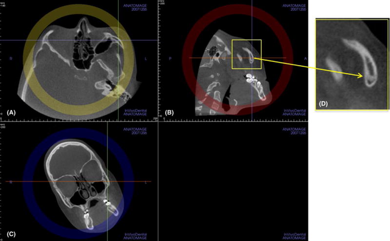
FIGURE 7.

Proper radiographic interpretation of the maturational stage of the inferior portion of the ZMS requires rolling the patient’s head in a counterclockwise direction in the coronal view (C) until the inferior portion of the ZMS is visualized properly in the sagittal view (B). (D) Close-up view of the inferior portion of the left ZMS [Colour figure can be viewed at wileyonlinelibrary.com]
