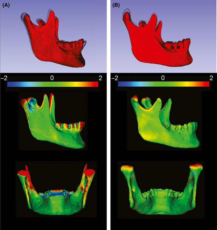
FIGURE 2.

Semi-transparent overlays of the 3D models (T0, red; and T1, black mesh), and closest point colour maps in the qualitative assessment of the condylar growth (mandibular regional superimposition). A, Herbst appliance patient. B, Comparison group patient.
