
Fig. 2.
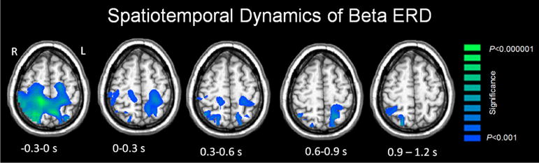
Statistical parametric maps (SPMs) showing the dynamics of beta oscillations (16–24 Hz) during the isometric knee extension motor task. The images have been thresholded at (p < 0.001, cluster-corrected) and are displayed following the radiological convention (R = L). As shown, there was a prominent beta ERD that was spread across the pre/postcentral gyri, SMA and parietal cortices during the motor planning stage (−0.3 to 0.0 s). The strength of the beta ERD diminished across all brain regions as children initiated the isometric knee extension force towards the displayed target (0.0–0.3 s), and this decline in beta ERD amplitude progressively continued throughout the isometric knee extension task (0.0–1.2 s)