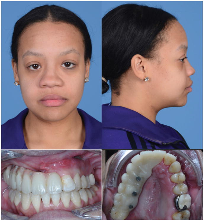
Figure 4.
Four month post-operative appearance. A – frontal and B – lateral views of patient following left hemi-LeFort I advancement, right maxillary reconstruction with free fibula flap and immediate placement of dental implants and prosthesis. C,D – Intra-oral photographs demonstrate correction of underjet and fistula. Posterior open bite on right to be corrected by repositioning of prosthesis after fibula healing.

