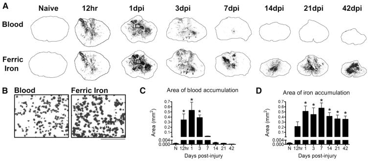
Fig. 1.
Iron and red blood cells infiltrate the lesion by 12 h post-injury and iron remains elevated chronically. A) Blood from intraspinal hemorrhage is evident in the injury site by 12 h post-injury and is markedly reduced after 3 days post-injury (dpi). Adjacent sections labeled with the Perls stain to visualize ferric iron reveal a close juxtaposition of iron and blood components through 3 dpi. Thereafter, blood is minimal but dense intraspinal iron remains within the tissue. B) High magnification of boxes from 1 dpi sections in A shows that the localization of iron is morphologically similar to that of infiltrating cells. C) Quantification of the area of blood in cross-sections from each time point shows that blood is significantly elevated for 3 dpi in the injured spinal cord. D) Quantification of Perls reactivity shows significant elevation of intraspinal iron by 12 h post-injury that is maintained for at least 42 dpi. C: *p < 0.05 vs. Naive. D: *p < 0.05 vs. Naive.

