
Figure 1. Transmission electronic micrographs of enterocytes during active dietary fat absorption.
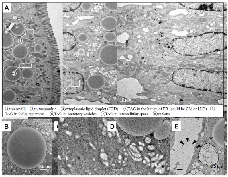
(A) An ultrastructural overview of enterocytes from the jejunum section of a wild-type C57BL6 male mouse, two hours after an oral gavage of 200 μl olive oil. At this time, TAG is directed to one of the subcellular pools and either stored in CLDs ➂ or packaged in CMs for secretion. CMs are synthesized and enlarged in the lumen of ER ➃, then transported to the cis-Golgi ➄ and Golgi-derived secretory vesicles ➅ for secretion from enterocytes ➆. (B) CLDs have cores of neutral lipids (TAGs and CEs) surrounded by a phospholipid membrane monolayer. (C) TAG is present within a phospholipid membrane bilayer of smooth ER. (D) TAGs are present within the Golgi apparatus. (E) CMs are carried by secretory vesicles (black arrows) and eventually secreted into the intercellular space (asterisk).