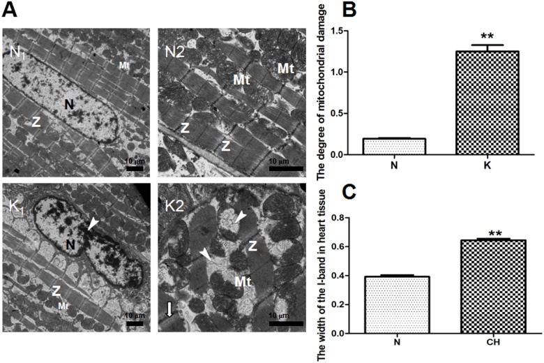
Fig 1. Heart tissue ultrastructure of the N and K groups by transmission electron microscope (TEM) and myocardial cell quantitative analysis of ultrastructural damege effects of kisspeptin-10 on mitochondria from group K and group N.
A: (N1) (N×1.2 k) The normal cardiomyocyte nuclei (N) and clear myocardial Z-band (Z) from the N group. (N2) (N×3.0 k) Normal mitochondria (Mt) from the N group. (K1) (K×1.2k) Margination of nuclear chromatin (↓) in the K group. (K2) (K×3.0 k) Injured mitochondrial cristae(▼) in the K group. B: The degree of mitochondrial damage. N: 0.19 ± 0.125, K: 1.25 ± 0.108. C: The width of the I-band in heart tissue. N: 0.393 ± 0.01 μm, K: 0.645 ± 0.01 μm, ** P < 0.01.

