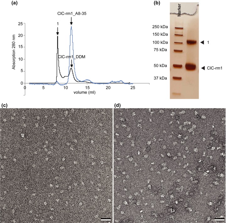
Fig 5. ClC-rm1 purified in DDM and exchanged with amphipol A8-35.
(a) Elution profile of ClC-rm1 in DDM from Superdex 200 SEC column (black line). SEC profile showed a large void volume peak of ClC-rm1 in DDM (labeled as 1) as well as a peak corresponding to the ClC-rm1 in DDM. ClC-rm1 in A8-35 has a symmetric peak with a very small void volume peak (highlighted in blue). (b) Silver stained SDS-PAGE gel of ClC-rm1 in A8-35 after SEC. Lane 1: molecular mass marker. Lane 2: ClC-rm1 in A8-35, ClC-rm1 monomer band at ~50 kDa with only one additional band (labeled as 1). (c) Micrograph of negatively stained ClC-rm1 in DDM used as initial materal for the A8-35 exchange experiment. (d) Micrograph of negatively stained ClC-rm1 in A8-35. Scale bar 20 nm.

