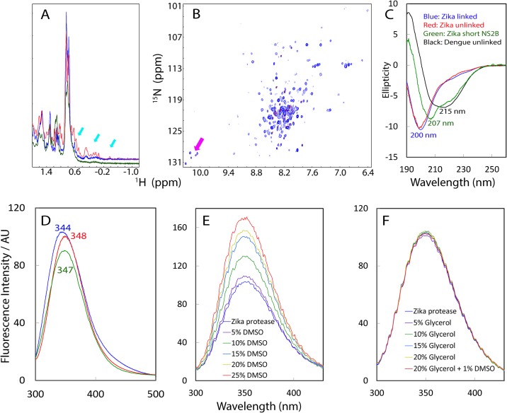
Fig 1. Biophysical characterization of Zika NS2B-NS3pro complexes.
(A) One-dimensional 1H NMR spectra over -1.2–1.8 ppm of linked (blue) and unlinked (red) Zika NS2B-NS3pro, as well as NS2B (48–74)-NS3pro (green) complexes at a protein concentration of 30 μM. Cyan arrows are used to indicate the very up-field peaks. (B) Superimposition of 1H-15N HSQC spectra of 15N-labeled unlinked (blue) and linked (red) NS2B-NS3pro complexes at a protein concentration of 30 μM. NMR spectra were acquired at 25°C in 10 mM sodium phosphate buffer at pH 6.5. Pink arrows are used to indicate five HSQC peaks for NS2B-Trp61, as well as NS3pro-Trp50, NS3pro-Trp69, NS3pro-Trp83 and NS3pro-Trp89 side chains only observed for the unlinked complex. (C) Far-UV CD spectra of linked (blue) and unlinked (red) Zika NS2B-NS3pro, as well as NS2B (48–74)-NS3pro (green) complexes at a protein concentration of 10 μM, together with that of Dengue-2 NS2B-NS3pro complex previously obtained (black) (ref. 12). (D) Emission spectra of the intrinsic UV fluorescence of linked (blue) and unlinked (red) Zika NS2B-NS3pro, as well as NS2B (48–74)-NS3pro (green) complexes at a protein concentration of 10 μM. (E) Emission spectra of the intrinsic UV fluorescence of unlinked Zika NS2B-NS3pro in 50 mM Tris-HCl buffer at pH 8.5, in the presence of DMSO at different concentrations. (F) Emission spectra of the intrinsic UV fluorescence of unlinked Zika NS2B-NS3pro in 50 mM Tris-HCl buffer at pH 8.5, in the presence of Glycerol as well as DMSO at different concentrations.

