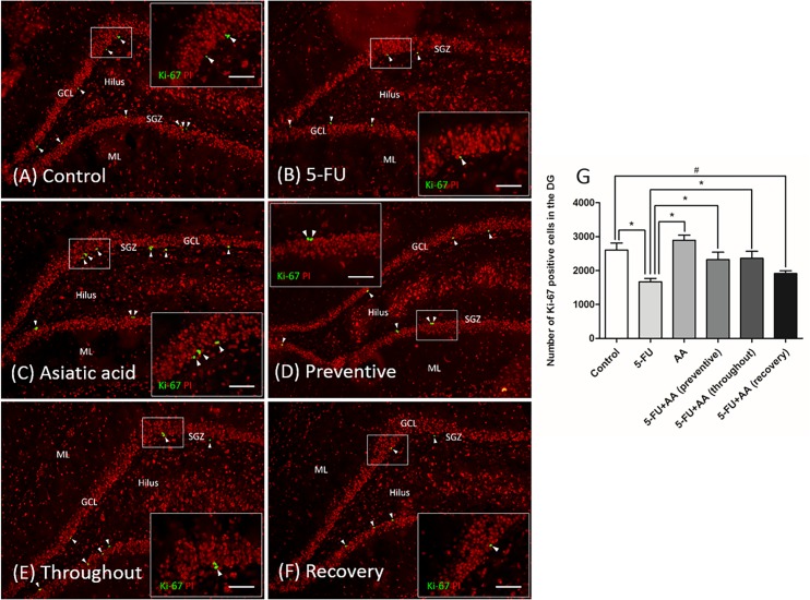
Fig 3. Effect of 5-FU and AA on proliferating cell counts in the dentate gyrus.
Representative images of Ki-67 positive cells in the dentate gyrus in each group (A-F). Ki-67 positive cells were stained green in the subgranular zone (SGZ) of the dentate gyrus. Section were counterstained with red nuclear dye, propidium iodide: PI. Arrowheads indicate Ki-67 positive cells in the the dentate gyrus (scale bars: 100 μm). Inserted images show Ki-67 immunostaining under high magnification (scale bar: 50 μm). Mean Ki-67 positive cell counts of the 5-FU group were significantly lower than the control, AA, and 5-FU+AA (preventive and throughout) groups (*p<0.05, G). Inaddition, Ki-67 positive cell number in the 5-FU+AA (recovery) group were significantly lower than in the control group (#p<0.05, G). ML: molecular layer, GCL: granule cell layer.

