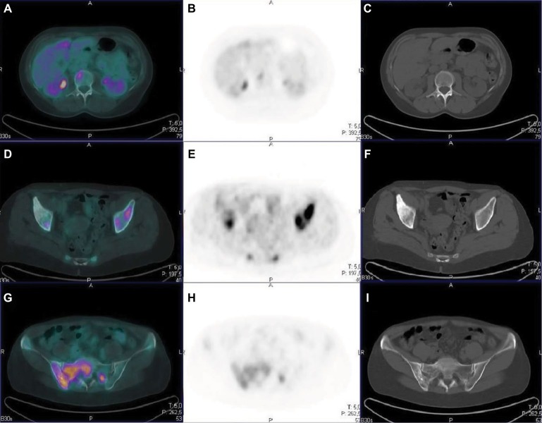
Figure 2.
Comparison between PET and CT in different types of bone lesions.
Notes: A–C: vertebral lesion with high FDG uptake in the absence of structural lesion on CT (metastasis in the bone marrow); D–F: CT sclerotic lesion in the right iliac bone with no concentration of FDG (likely a lesion with low cellularity); G–I: mixed bone lesion in the right sacroiliac bone on CT images markedly positive on the FDG PET scan.
Abbreviations: CT, computed tomography; FDG, fluorodeoxyglucose; PET, positron emission tomography.

