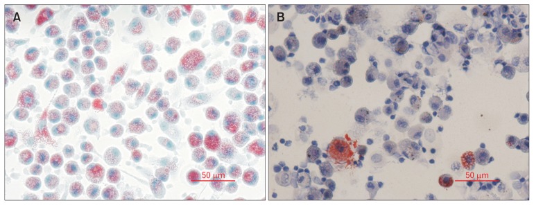
Figure 1.
Primary alveolar macrophages stained with Oil Red O and Hematoxylin. Bronchoalveolar lavage samples were processed and Cytospun onto glass slides. Cells were stained for lipids using Oil Red O and counterstained with Mayer’s Hematoxylin. Lipid droplets stained vivid red and were easily distinguishable against the purple counterstain. Patients showed a high degree of variability of lipid accumulation. Patient (A) shows many cells with lipid accumulation whilst patient (B) shows only a few cells with lipid accumulation (bar = 50 μm).

