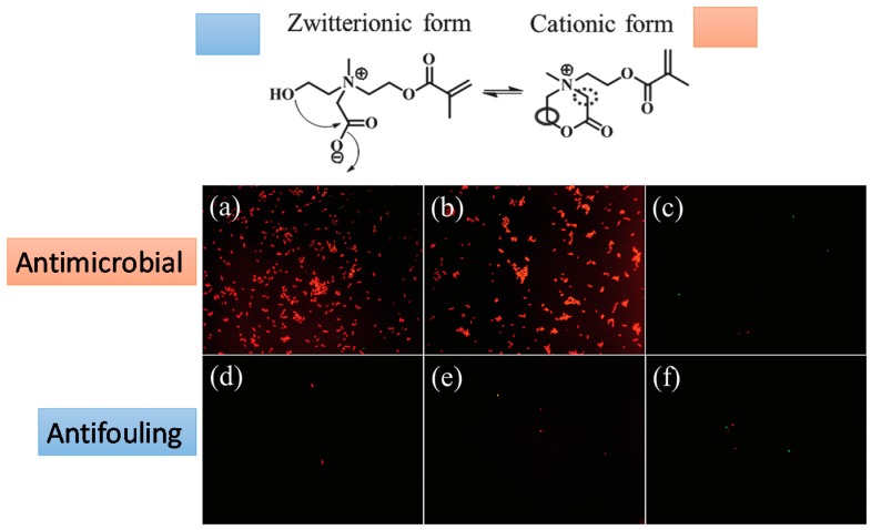
Figure 14.
Fluorescence microscopy images of bacterial attachment on pCBOH1 in cationic form (a); pCBOH2 in cationic form (b); and poly(carboxybetaine methacrylate) pCBMA (c) hydrogels before hydrolysis and on pCBOH1 in zwitterionic form (d); pCBOH2 in zwitterionic form (e); and pCBMA (f) hydrogels after 16 h of hydrolysis in PBS. Cells with damaged cytoplasmic membrane are in red, and cells with intact cytoplasm membrane are in green. Reproduced with permission from [40].

