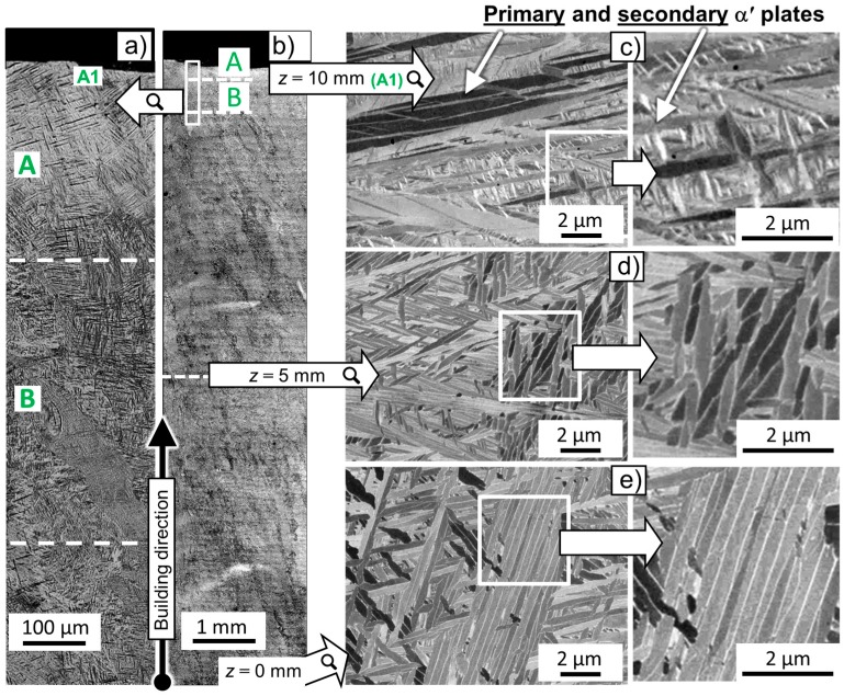
Figure 1.
Light optical microscopy (LOM) of (a) the upper region and of (b) the entire central cross section of a Ti-64 sample along the SLM building direction. Backscattered electron mode-scanning electron microscopy (BSE-SEM) images are shown, corresponding to selected regions of the microstructure for (c) z ~ 10 mm (top); (d) z ~ 5 mm (center) and (e) z ~ 0 mm (bottom).

