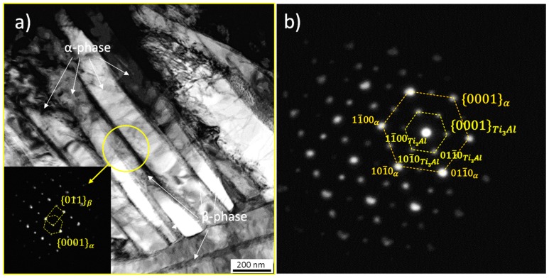
Figure 2.
(a) Bright field transmission electron microscopy (TEM) image showing the ultrafine α + β microstructure formed at the center of a Ti-64 SLM sample (see z ~ 5 mm in Figure 1). The inset shows the Burger’s orientation relationship {011}β // {0001}α between crystal lattices; (b) Selected area electron diffraction pattern revealing the presence of α2-Ti3Al within an α lamella.

