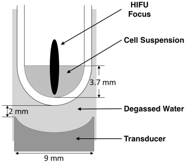
Figure 1.
Experimental setup showing one of the four transducers and sample wells. Each transducer is approximately the diameter of a microplate well, and situated 2 mm below the well bottom, with degassed water as the coupling medium. The bacterial sample suspension filled the well to a height of 3.7 mm. The free-field focal spot is overlaid in this drawing, but is not to scale.

