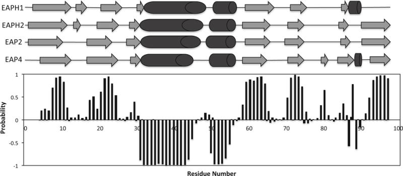
Fig. 2.

Secondary structure prediction for the Eap4 domain based on the TALOS+ program using obtained chemical shift values. β-strand probabilities are given by positive values, α-helices are given by negative values, and loop regions are given by values approximately from −0.3 to 0.3. Shown in the top portion are the secondary structure topology obtained from the published crystal structure of Eap2 (Geisbrecht et al. 2005) and the TALOS+ prediction of Eap4 with α-helices shown as cylinders and β-sheets shown as arrows. The predicted secondary structure of Eap4 shows very similar topology to the other three EAP domains whose crystal structures are available
