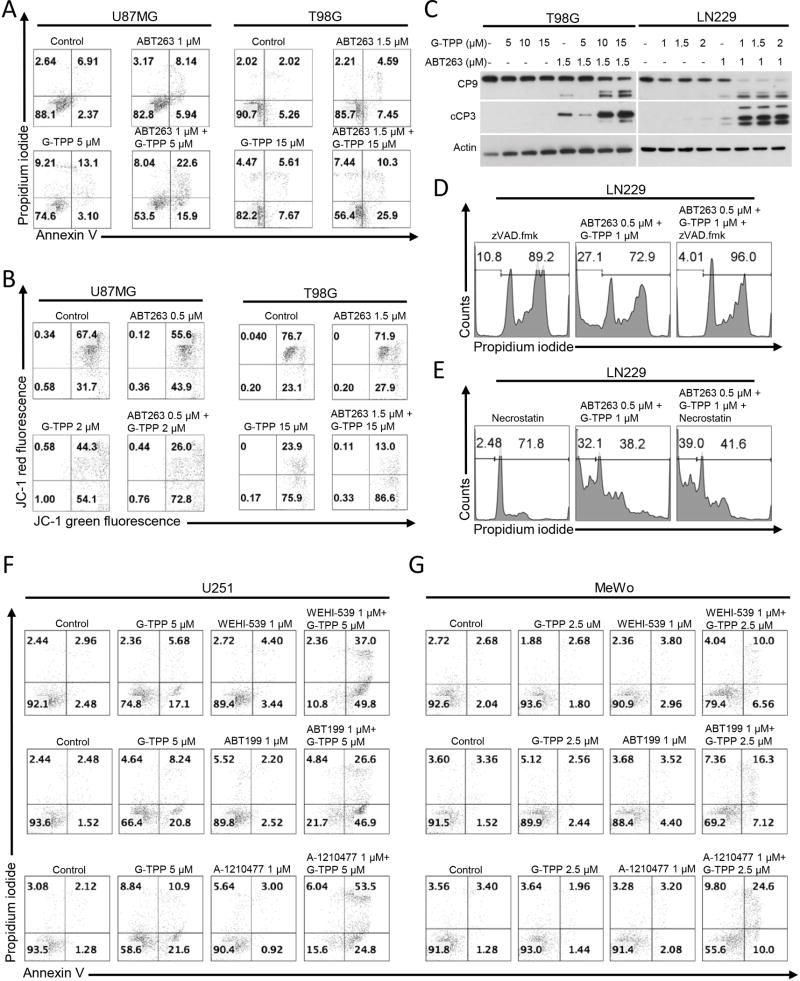
Figure 2.
Combined treatment with G-TPP and broad or selective BH3-mimetics yields enhanced induction of apoptosis. A, Representative histograms of U87MG and T98G glioblastoma cells treated with solvent, ABT263, G-TPP or the combination as indicated for 48h prior to staining for Annexin V and propidium iodide and flowcytometric analysis. B, Representative histograms of U87MG and T98G glioblastoma cells that were treated for 24 h with G-TPP, ABT263, both or solvent prior to staining with JC-1 and flow cytometric analysis. C, T98G and LN229 glioblastoma cells were treated for 7 h with G-TPP, ABT263, both agents and solvent under serum starvation. Whole-cell extracts were examined by Western blot analysis for caspase 9 (CP9) and cleaved caspase 3 (cCP3). Actin Western blot analysis was performed to confirm equal protein loading. D, LN229 cells were treated with the combination of ABT263 and G-TPP as indicated in the presence or absence of the pan-caspase inhibitor zVAD.fmk (20µM). Staining for propidium iodide and flowcytometric analysis was performed to determine the fraction of subG1 cells. E, LN229 cells were treated with the combination of ABT263 and G-TPP as indicated in the presence or absence of necrostatin (20µM). Staining for propidium iodide and flowcytometric analysis was performed to determine the fraction of subG1 cells. F–G, U251 glioblastoma (F) or MeWo melanoma (G) cells were treated with selective BH-3 mimetics, G-TPP or the combination of both for 24 h (U251) or 48 h (MeWo). Thereafter, cells were stained with annexin V and propidium iodide and analyzed by flow cytometry. Shown are representative flow plots.

