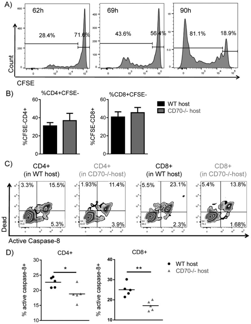
Figure 7. Donor T cells in CD70-/- hosts have similar proliferation, but significantly decreased caspase 8-dependent activation induced cell death.

(A-B) WT or CD70-/- C57BL/6 mice received 965 cGy irradiation on day -1 and were subsequently transplanted on day 0 with 4×10⁶ BALB/c BM + 5×10⁶ CFSE labeled PanT. (A) Represented histograms of CFSE dilution in donor-derived live H-2Kd+CD8+ T cells in WT hosts at the indicated hours after allo-HCT. (B) 69 hours after allo-HCT, CFSE dilution was assessed in donor-derived live H-2Kd+CD4+ and H-2Kd+CD8+ T cells. Summary data from 4 mice of each genotype. (C-D) WT or CD70-/- C57BL/6 mice received 965 cGy irradiation on day -1 and were subsequently transplanted day 0 with 4×10⁶ BALB/c BM + 5×10⁶ PanT. 4 days post allo-HCT spleens were harvested and active caspase-8 was evaluated in H-2Kd+H-2Kb-TCRβ+CD4+ and H-2Kd+H-2Kb-TCRβ+CD8+ T cells. (C) Flow diagrams depicting T cell expression of live/dead viability dye versus active caspase-8 in WT and CD70-/- hosts. (D) Summary data from 5 mice of each genotype. Representative data from one of three individual experiments is shown. Data were analyzed by student's t test. *P<0.05; **P<0.01.
