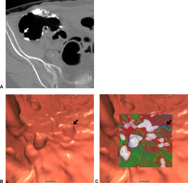
Figure 2. Soft tissue polyp in the setting of abundant tagged solid stool related to “minimal preparation” with 40% barium.
2D CTC image (A) shows abundant densely tagged stool in the right colon. A homogeneous soft tissue lesion (arrow), which proved to be a true polyp, is seen adjacent to polypoid tagged stool. 3D endoluminal images without (B) and with (C) translucency rendering show multiple polyp candidates, but only one true soft tissue lesion (arrows). This degree of residual solid stool makes polyp detection very difficult, even when tagged. In addition, we recommend the use of 2% barium over 40%, which is too dense.

