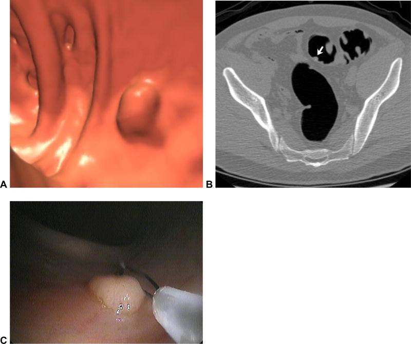
Figure 7. Primary 3D versus primary 2D polyp detection at CTC.
3D (A) and 2D (B) images from CTC show a 9-mm polyp (arrow) in the sigmoid colon. The polyp is easy to distinguish from adjacent folds on 3D, but the task is much more difficult on the 2D view alone (without the arrow, at least). The lesion was confirmed at same-day OC (C) and proved to be hyperplastic.

