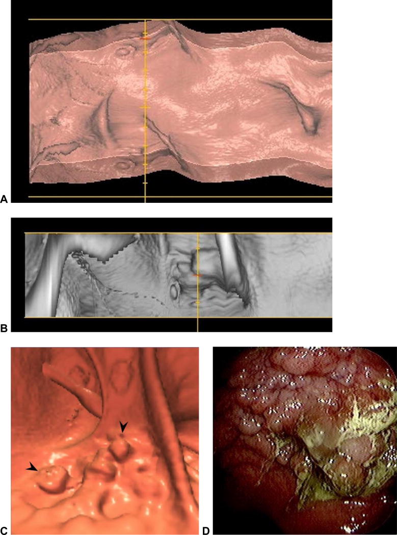
Figure 8. Spatial distortion obscuring a large rectal mass at CTC.
3D virtual dissection with 360? display (plus overlap) (A) shows subtle irregularity where the yellow line intersects. The same area in the rectum is shown on the 120? strip (B), which begins to show a mass lesion. On the standard 3D endoluminal view (C), the lobulated carpet lesion (arrowheads) between rectal folds is more apparent. This proved to be a large villous adenoma with high-grade dysplasia at OC (D).

