
Figure 5. Extensive interactions between Cdt1 and MCM hexamer.
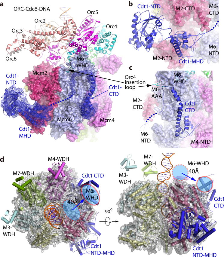
(a) OCCM structure with Cdt1 electron density shown in blue mesh. The CTD of Cdt1 locates between Mcm6 and Mcm4, over 60 Å away from the NTD and MHD of Cdt1. (b) Zoomed view of the Cdt1 NTD and MHD showing their interactions with Mcm2 and Mcm6. (c) Zoomed view showing the Cdt1 CTD interacting with Mcm6 WHD. The dotted blue line in (a-c) indicates a flexible loop connecting Cdt1 MHD and CTD. (d) The top view (left) and front side view (right) of Mcm2-7 structure in cartoon and semi-transparent surface view. The red oval marks Mcm6-WHD in OCCM, and the dashed red oval the position of Mcm6-WHD in CMG helicase. The blue arrow shows the displacement of Mcm6 WHD in OCCM due to interaction with Cdt1 CTD. Such displacement forms an unobstructed Mcm2-7 C-terminal face for binding with ORC-Cdc6.