. 2017 Jun 26;6:e25736. doi: 10.7554/eLife.25736

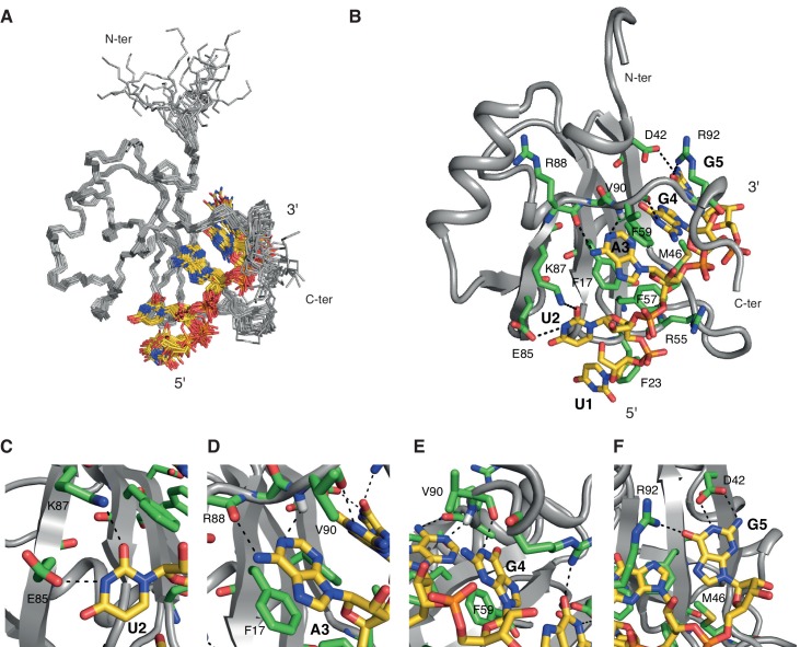
Figure 1. Overview of the solution structure of hnRNP A1 RRM1 bound to 5´-UUAGGUC-3´ RNA.

(A) NMR ensemble. Overlay of the 20 final structures superimposed on the backbone of the structured part (11-92) and represented as a ribbon trace (N, Cα, C´). The not-well-defined nucleotides U6 and C7 are omitted for a better overview. The RNA is shown in stick representation with the carbon atoms in yellow, nitrogen in blue, phosphate in orange and oxygen in red. (B) Cartoon drawing of a representative structure of the NMR ensemble. Residues with important roles in RNA binding are shown as sticks, with the carbon atom in green. All other atoms have the same color code as in panel A. (C–F) Close-up views of each single nucleotide recognition by hnRNP A1 RRM1. Representation and colors are similar to panel B. Residues with important roles in RNA binding are labeled.

DOI: http://dx.doi.org/10.7554/eLife.25736.002

Figure 1.

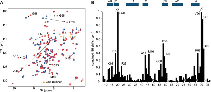
Figure 1—figure supplement 1. Overview of the titration of 5´-UUAGGUC-3´to hnRNP A1 RRM1.

Figure 1—figure supplement 1.

(A) NMR titration of hnRNP A1 RRM1 with the 5´-UUAGGUC-3´ RNA. Overlay of a 2D (15N,1H)-HSQC of the free form in blue and the bound form at a 1:1 ratio in red. Aliased peaks are colored in cyan (free) and orange (bound). Residues with large chemical shift perturbations are highlighted with an arrow. (B) Plot of the combined backbone NH chemical shift perturbation of hnRNP A1 RRM1 upon binding to the 5´-UUAGGUC-3´ RNA. Residues with large chemical shift perturbations are labeled on the histogram. (P) corresponds to proline residues and (*) to missing amide signals in either the free or bound form. Secondary structure elements are depicted above the histogram.
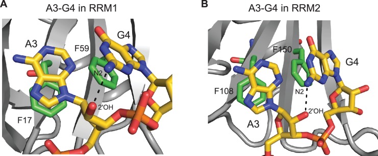
Figure 1—figure supplement 2. Intramolecular hydrogen bond within the AG dinucleotide constituting the core of the hnRNP A1 RRMs binding motif.

Figure 1—figure supplement 2.

Intramolecular hydrogen bond within the AG dinucleotide in the structure of (A) hnRNP A1 RRM1 bound to 5´-UUAGGUC-3´ RNA; or (B) hnRNP A1 RRM2 bound to 5´-UCAGUU-3´ RNA. The intramolecular hydrogen bond occurs between the N2 exocyclic amino-group of G4 and the 2´-hydroxyl group of the A3 ribose. This intramolecular hydrogen bond is specific of RNA oligonucleotides and is therefore not found in the structures of UP1 bound to DNA oligonucleotides.