. 2017 Jun 26;6:e25736. doi: 10.7554/eLife.25736

Figure 5. SMN splicing repression depends on hnRNP A1 organization and the bipartite ISS-N1 motif.

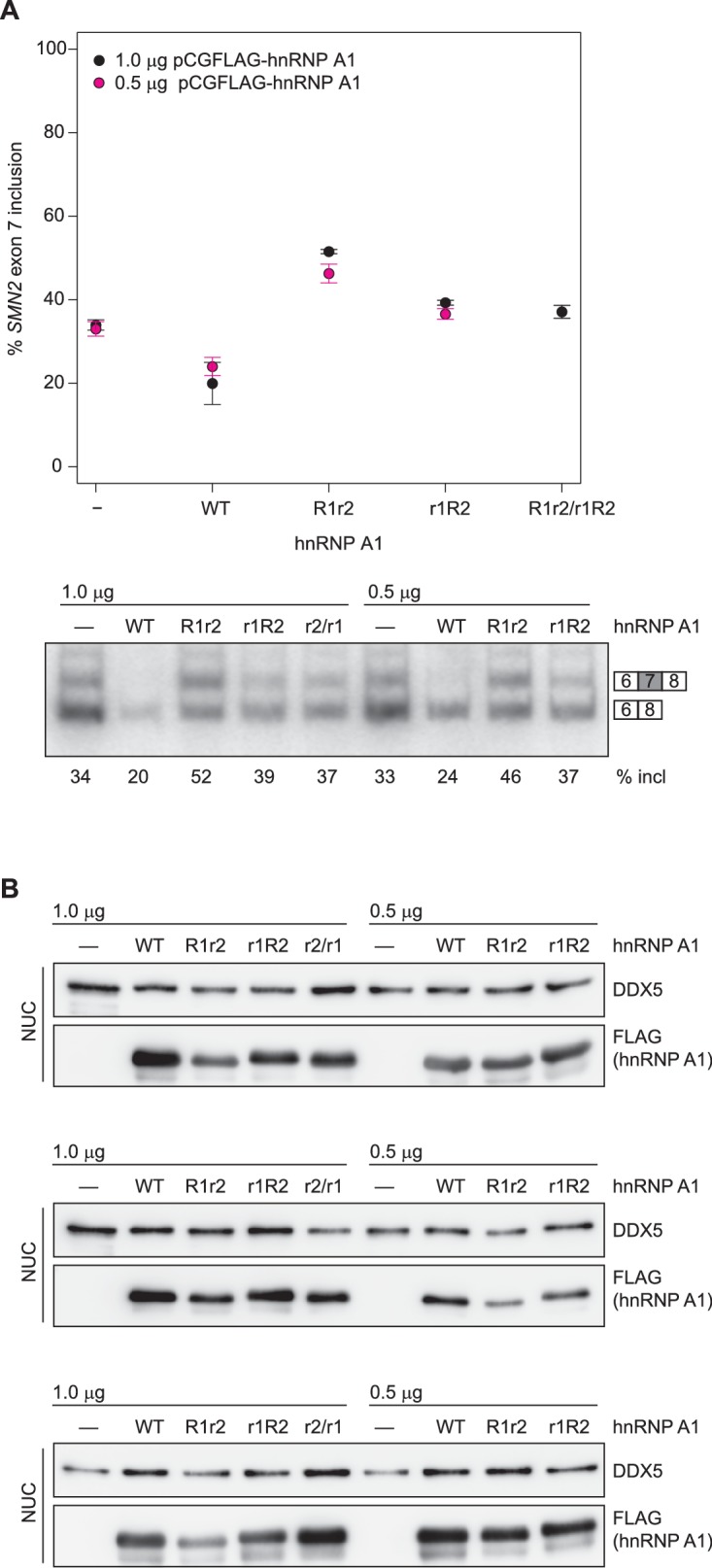
(A) Data from alternative splicing assay in HEK293T cells. Top panel, quantification of exon 7 inclusion in SMN1 upon overexpression of wild-type or mutant hnRNP A1. Bottom panel, PAGE with PCR products used for quantification. (B) Western Blot of nuclear extracts used in A. (C, D) Quantification of exon 7 inclusion in SMN2 in relation to overexpression of wild-type or mutant hnRNP A1 (C) or to mutation of the hnRNP A1 binding sites in the ISS-N1 (D). Both figures (C) and (D) contain are different representations of the same data. (E) Radioautography of PAGE with PCR products used for quantification in (C, D).The separation between two individual gels is indicated by an arrow. (F) Quantification of exon 7 inclusion in SMN2 in relation to the spacer length in-between the two hnRNP A1 binding sites within the ISS-N1. The wild-type ISS-N1 has a spacer length of 9 nt. (G) Radioautography of PAGE with PCR products used for quantification in (F). The separation between two individual gels is indicated by an arrow. Data information: All data points represent the mean of the biological replicates. Error bars correspond to the S.E.M. (B) **p=0.00113 (INT2), p=0.00360 (INT3), ***p<0.001; (C) ***p<0.001; (D) *p<0.5, **p<0.01, ***p<0.001; (F) *p=0.01176, **p=0.00287. All tested with one-way ANOVA (WT = 0), for panel (C, D) SMN2 wt co-transfections n = 4, otherwise n = 3. The mean of the replicate is always given below the corresponding PAGE for a better readability. If not otherwise indicated 0.5 µg hnRNP A1 were co-transfected with 1 µg of the mini-gene.

DOI: http://dx.doi.org/10.7554/eLife.25736.022

Figure 5—source data 1. PAGE quantification for SMN exon 7 inclusion (%).
DOI: 10.7554/eLife.25736.023

Figure 5.

Figure 5—figure supplement 1. Protein mutants disrupt the inter-RRM interface.

Figure 5—figure supplement 1.

(A) Overlay of (15N,1H)-HSQCs of wild-type UP1 (in gray) and of UP1-INT1 (D157K +R75A) double-mutant (in blue). (B) Overlay of (15N,1H)-HSQCs of wild-type UP1 (in gray) and of UP1-INT2 (D155R +D157K) double-mutant (in blue). (C) Overlay of (15N,1H)-HSQCs of wild-type UP1 (in gray) and of UP1-INT3 (D155R +D157K+I164A) triple-mutant (in blue). The mutants are designed to disrupt the inter-RRM interface by removing the two salt-bridges at the interface, namely the R75:D155 and the R88:D157 salt-bridges. (D) 15N T1, T2 and overall correlation time (τC) of UP1 wild-type (UP1-WT) and UP1-INT1, UP1-INT2 and UP1-INT3 inter-RRM interface mutants. Transverse relaxation time T2 is increased in UP1-INTs mutants as compared with UP1-WT. This indicates a faster tumbling of the single RRMs in the double mutant than tumbling as one unit in the wild-type UP1, confirming that the RRM-RRM interaction is perturbed. The UP1-INT2 mutant appears to be the most affected mutant.
Figure 5—figure supplement 2. No dose dependency of hnRNP A1 overexpression.

Figure 5—figure supplement 2.

(A) Data from alternative splicing assay in HEK293T cells. Quantification of exon 7 inclusion in SMN1 upon overexpression of wild-type or mutant hnRNP A1. 0.5 µg, 1 µg, or 2 µg pCGFLAG-hnRNP A1 WT/mutant were cotransfected with 1 µg pSMN1. SMN1 exon 7 inclusion remains stable regardless of increased hnRNP A1 overexpression. (B) Western Blots of nuclear extracts used in (A).
Figure 5—figure supplement 3. Western blots for SMN2 exon 7 splicing experiments in HEK293T cells.

Figure 5—figure supplement 3.

(A) Western Blots of nuclear extracts used in Figure 5C–E. (B) Western Blots of nuclear extracts used in Figure 5F,G.
Figure 5—figure supplement 4. hnRNP A1 RRM mutants can not complement each other in SMN2 splicing assay.

Figure 5—figure supplement 4.

(A) Data from alternative splicing assay in HEK293T cells. Quantification of exon 7 inclusion in SMN2 upon overexpression of wild-type or mutant hnRNP A1. HnRNP A1 R1r2 and r1R2 were independently as well as co-transfected. The mutants cannot complement each other as evidenced by same SMN exon 7 inclusion rate as for independent transfection of hnRNP A1 r1R2. (B) Western Blots of nuclear extracts used in (A).