
Figure 6. HGF sustains glycolysis in low glucose by promoting GLUT1-mediated glucose uptake.
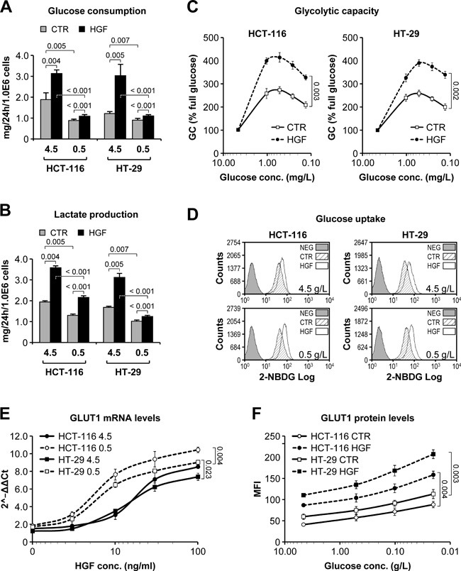
A. Glucose consumption by HCT-116 and HT-29 cells grown in high (4.5g/L) or low (0.5g/L) glucose-containing medium and in the absence or presence of HGF was determined over a period of 24 hours. B. Same as in A but measuring lactate production. C. Glycolytic capacity (GC) of HCT-116 and HT-29 cells pre-incubated in progressively lower glucose concentrations and in the absence or presence of HGF was determined by Seahorse technology. GC is expressed as % relative to full glucose. D. Incorporation of the fluorescent glucose analogue 2-NBDG was determined by flow cytometry in HCT-116 and HT-29 cells pre-incubated in high (4.5g/L) or low (0.5g/L) glucose-containing medium and in the absence or presence of HGF. NEG, negative control. E. GLUT1 mRNA levels were determined by RT-PCR analysis of HCT-116 and HT-29 cells grown in high (4.5g/L) or low (0.5g/L) glucose-containing medium and in the presence of increasing HGF concentrations. Data are expressed as 2−ΔΔCt compared to cells without HGF. F. GLUT1 protein levels were determined by flow cytometry analysis of HCT-116 and HT-29 cells grown in decreasing glucose concentrations and in the absence or presence of HGF. MFI, mean fluorescence intensity. Values represent mean ± SEM. Statistical significance was calculated using a Student's T-test (n = 3).