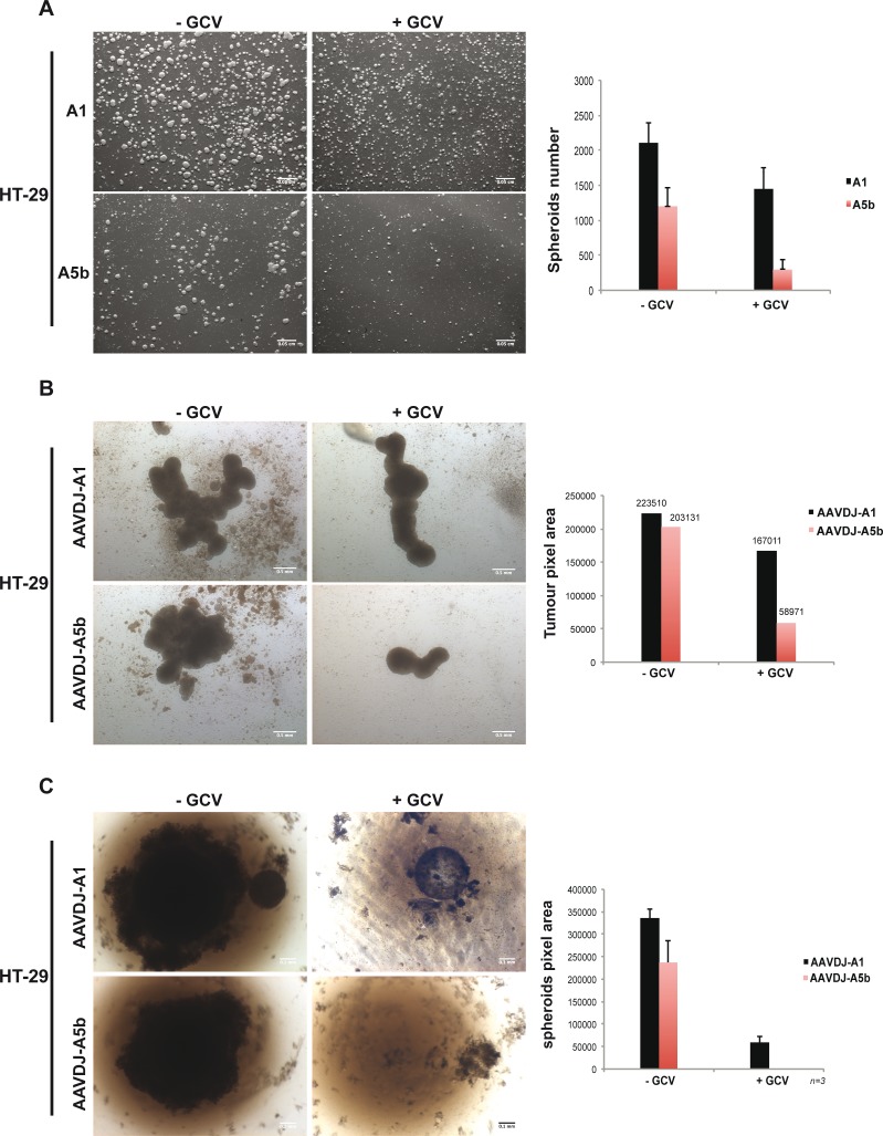
Figure 5. Targeting spheroid growth of HT-29 cells.
A. HT-29 cells were transfected twice as described above and 48 hours post transfection, 106 cells were cultured in bacterial petri dishes, with or without GCV. One-week later, spheroids were imaged and counted. Scale bar; 50 μm. B. 105 HT-29 cells were transferred onto agarose-coated 12-well plates and infected at 107 MOI and GCV was added after 4 hours. Two weeks later spheroids were imaged and measured. Scale bar; 50 μm. C. Similar to MCF-7 cells above, HT-29 cells were grown as single spheroids in a 96 well plate with 2 cycles of infection and spheroids were imaged and measured after 1 month of growth. Scale bar; 50 μm.

