
Figure 4. Laminarin-promoted maturation of drLNs and DCs in the tumor microenvironment.
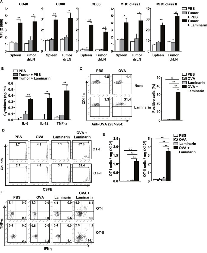
C57BL/6 mice were inoculated s.c. with 1 × 10⁶ B16 melanoma cells or B16-OVA cells. Fifteen days after tumor injection, the mice were treated with PBS and 25 mg/kg of laminarin for 24 hours. (A) MFI of CD40, CD80, CD86, and MHC classes I and II levels were measured in the spleen and tumor drLNs and DCs. (B) Concentrations of IL-6, IL-12p40, and TNF-α in the mice sera. (C) Surface OVA peptide (257-264) presentation was measured in the tumor drLNs and DCs. (D) CFSE-labeled OT-I and OT-II T cell proliferation in B16-OVA tumor-bearing CD45.1 congenic mice were analyzed using flow cytometry. (E) The means of the absolute numbers of OT-I (left panel) and OT-II (right panel) cells in the tumor. (F) Percentage of IFN-γ+ and TNF-α+ cells in tumor-infiltrated OT-I and OT-II cells. All data are representative of or the average of analyses of six independent samples (two mice per experiment, for a total of three independent experiments). **p < 0.01, *p < 0.05.