
Figure 6. Treatment with laminarin and OVA inhibited liver metastasis of B16-OVA tumor cells.
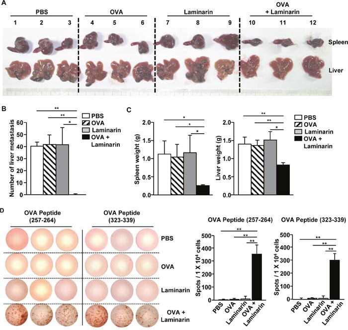
C57BL/6 mice were treated i.v. with PBS, 50 μg of OVA, 25 mg/kg of laminarin, and the combination of laminarin and OVA. Three days after the treatment, the mice were inoculated i.s. with B16-OVA melanoma cells. On day 3 after the B16-OVA cell challenge, the mice again received the same amount of the laminarin and OVA treatment. (A) The size of the tumor masses in the spleens and liver metastasis of B16-OVA cells on day 14 after tumor injection. (B) The mean of the absolute number of B16-OVA metastasis in the livers. (C) Mean weights of spleens (left panel) and livers (right). (D) Splenocytes were harvested without tumor cells. OVA peptide-specific IFN-γ production in the splenocytes was analyzed using ELISPOT analysis (left panel). The mean number of IFN-γ positive spots (right panel). All data are representative of or the average of analyses of six independent samples (three mice per experiment, for a total of two independent experiments). *p<0.01, **p < 0.01.