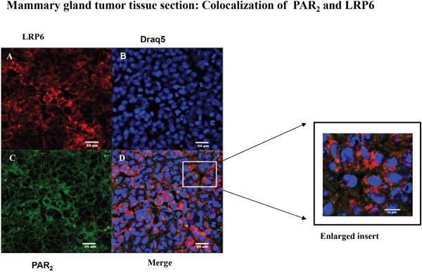
Figure 7. Colocalization between LRP6 and PAR2 in breast mouse cancer tumor biopsies: confocal analysis.

Confocal immunohistostaining was carried out on PAR2-induced mouse orthotopic mammary gland tumors. Application of anti-LRP6 (A; red) and anti-PAR2 (C; green) Abs was followed by appropriate Cy2- and Cy3-conjugated IgG secondary antibodies. The cell nuclei were visualized by DRAQ5 (B; blue). High expression levels were observed in both LRP6 and PAR2. Merge analyses indicate co-localization between LRP6 and PAR2 (D; orange). Data shown are representative of three independent experiment. Magnification x40.
