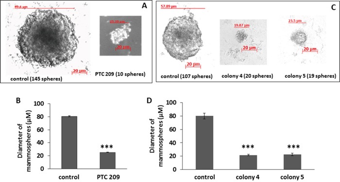
Figure 3. FMMC 419II cells treated with 2 μM PTC 209 or transfected with Bmi1 shRNA display significantly lower mammosphere formation potential.

Treated cells were plated at a concentration of 1 cell/μl in an ultra-low attachment 96-well plate in serum free conditions. Phase contrast images at 20X magnification were taken of mammospheres (“spheres”) that were formed after a 2 week incubation (A, C). The graphs (B, D) represent the mean ± S.E.M. of the mammosphere diameters. ***P <0.005.
