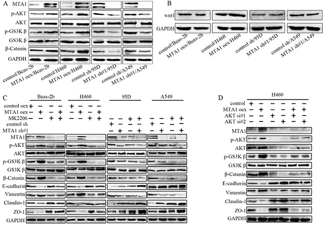
Figure 6. MTA1 promotes NSCLC cell EMT through the AKT/GSK3β/β-catenin signaling pathway.

(A) Western blot analysis of AKT, p-AKT, GSK-3β, p-GSK-3β, and β-catenin expression, (B) Western blot analysis of Wnt1. (C) Western blot analysis of MTA1, AKT, p-AKT, GSK-3β, p-GSK-3β, β-catenin, and EMT markers (E-cadherin, Vimentin, Claudin-1, and ZO-1). Cells were treated with 10 μM MK2206 for 24 h. (D) Western blot analysis of MTA1, AKT, p-AKT, GSK-3β, p-GSK-3β, β-catenin, and EMT markers (E-cadherin, Vimentin, Claudin-1, and ZO-1). H460 cells were transfected with MTA1-overexpression plasmid for 72 h, or transfected with siRNA for 48 h, or transfected with MTA1-overexpression plasmid for 24 h followed by siRNA for 48 h. GAPDH was used as a loading control. oex: overexpression; si#1: siRNA#1; si#2: siRNA#2.
