
Figure 1. CD47 is highly expressed in ovarian cancer and correlates with poor clinical outcome.
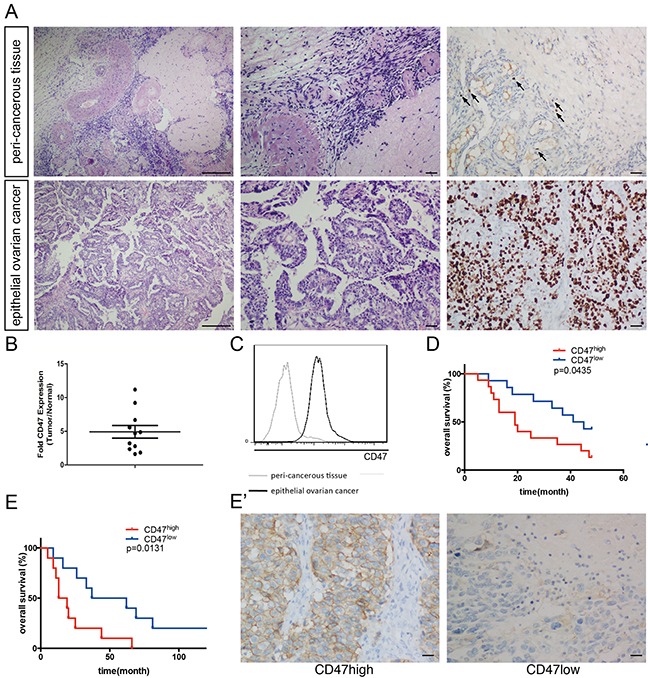
(A) Representative pathology of patients. Left: ×40 magnification H&E staining (scale bar = 250 μm); middle: ×100 magnification H&E staining (scale bar = 50 μm); right: ×100 magnification IHC staining of Ki-67 (scale bar = 50 μm). (B) CD47 expression in the epithelial cancer tissues and peri-cancerous tissues in a representative patient. (C) Relative CD47 expression in epithelial ovarian cancer tissues normalized to morphologically normal peri-cancerous tissues, measured by MFI fold change using flow-cytometry. (D) Kaplan-Meier survival curve of CD47high (n = 15) and CD47low (n = 14) patients in the high-grade serous ovarian cancer cohort (Log-rank test, *p < 0.05). (E) Ten-year survival curve of CD47high (n = 10) and CD47low (n = 10) patients in the high-grade serous ovarian cancer (Log-rank test, *p < 0.05). (E') Representative IHC staining of CD47 in the high-grade serous ovarian cancer cohort (scale bar = 50 μm).