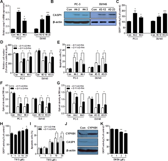
Figure 4. CASP1 is a functional target of CYP1B1.
(A–C) CASP1 mRNA (A), protein (B), and enzyme activity (C) in CYP1B1 shRNA stably expressing PCa cells. Levels were determined by qRT-PCR, Western blot and colorimetric assay, respectively. (D–G) Effect of CASP1 inhibition on the tumorigenicity of CYP1B1 shRNA stably expressing PCa cells. Cells were treated with Z-YVAD-fmk (100 μM) and proliferation (D), apoptotic cell death (E), migration (F), as well as invasion (G) were examined, respectively. (H) Effect of chemical inhibition of CYP1B1 on CASP1 activation. CASP1 activity was determined by colorimetric assay in PC-3 cells treated with the indicated concentration of TMS. (I) Effect of CASP1 inhibition on apoptotic cell death induced by chemical inhibition of CYP1B1. Apoptotic cell death was determined by flow cytometric analysis using double staining with Annexin V-FITC and 7-AAD in TMS-treated PC-3 cells with or without addition of Z-YVAD-fmk (100 μM). (J and K) Effect of CYP1B1 activation on CASP1 in RWPE-1 cells. CASP1 expression was determined by Western blot after CYP1B1 overexpression (J). CASP1 activity was examined by colorimetric assay in cells treated with the indicated concentration of DMBA (K). *p<0.05; **p<0.01; ***p<0.001.

