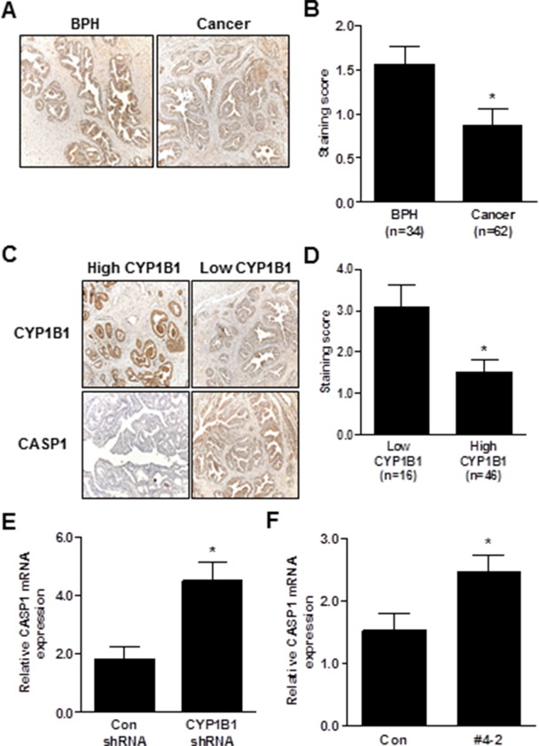
Figure 6. Inverse correlation between CYP1B1 and CASP1 expression.
(A and B) Immunohistochemical staining of CASP1 protein in PCa specimens. Representative images showing immunoreactive caspase-1 in BPH and cancer tissues (magnification: × 200) (A). Summary of CASP1 immunostaining score (B). (C and D) Immunohistochemical staining of CYP1B1 and CASP1 protein in prostate cancer specimens. Representative images showing inverse correlation of CYP1B1 and CASP1 in BPH and cancer tissues (magnification: × 200) (C). Summary of CASP1 immunostaining score (D). (E and F) Expression of CASP1 mRNA in tumors injected with CYP1B1 shRNA (E) and PC-3/CYP1B1 shRNA #4-2 xenografts (F). *p<0.05.

