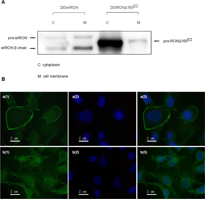
Figure 3. Cellular localization of RONΔ165E2.
(A) Using a Cell Surface Protein Isolation Kit, the cytoplasmic and cell-surface proteins were separated by SDS-PAGE and incubated with an antibody against RON (H160). In HEK293 wtRON cells, the pro-RON (180kD) and mature RON protein (150 kDa) both existed on the cell surface, while in HEK293 RONΔ165E2 cells, the variant protein was expressed in the cytoplasm. (B) Detection of RON expression by immunofluorescence staining in both HEK293 wtRON and HEK293 RONΔ165E2 cells. a(1), b(1) were respectively on behalf of the location about wtRON protein, RONΔ165E2 protein which were dyed green by FITC. a(2), b(2) nuclei were counterstained with DAPI (blue). a(3) was the merged picture of a(1) and a(2), while b(3) was the merged picture of b(1) and b(2).

