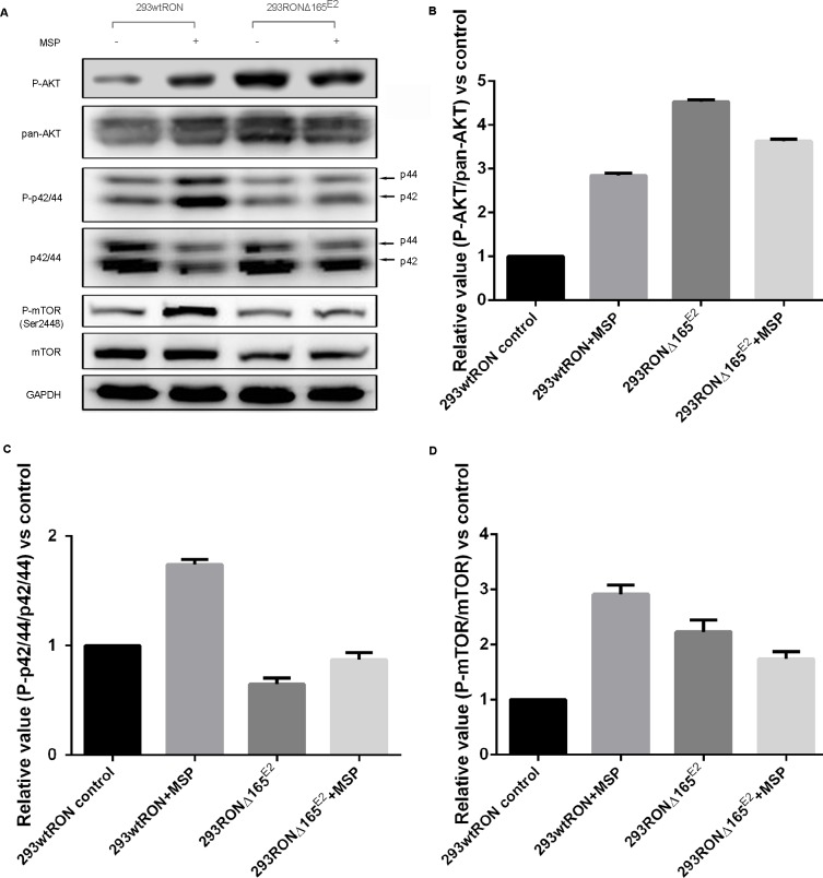
Figure 4. Constitutive activation of the PI3K/Akt pathway in 293 RONΔ165E2 cells.
(A) Western blotting results on the activation of signaling proteins involved in the PI3K/Akt and MAPK pathways. (B) The relative ratio of phosphorylated to total Akt expression in HEK293 wtRON cells was significant after MSP stimulation. While there was no meaningful change in the relative ratio of phosphorylated to total Akt expression in HEK293 RONΔ165E2 cells with or without MSP, it was significantly higher than that in HEK293 wtRON cells even with MSP stimulation (p < 0.05). (C) Relative value of p-p44/42/p44/42 in HEK293 wtRON cells was significantly increased after MSP stimulation. While there was no meaningful change in HEK293 RONΔ165E2 cells with or without MSP, it was significantly lower than that in HEK293 wtRON cells (p < 0.05). (D) Relative ratio of p-mTOR/mTOR was increased in HEK293 RONΔ165E2 cells compared with HEK293 wtRON cells (p < 0.05). Compared with HEK293 wtRON cells in the quiescent state, the level of the relative p-mTOR/mTOR ratio was significantly increased after they were treated by MSP. MSP had no effect on the level of mTOR activation in HEK293 RONΔ165E2 cells.

