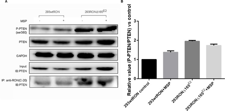
Figure 5. PTEN lipid phophatase activity was decreased in HEK293 RONΔ165E2 cells despite the upregulated PTEN protein expression.
(A) western blotting results using the antibodies against PTEN, P-PTEN(Ser380) indicated higher level in HEK293 RONΔ165E2 cells compared to the HEK293 wtRON cells, p < 0.05. After co-immunoprecipitation with the antibody RON(C-20), western blotting with the antibody specific to PTEN showed that the RONΔ165E2 protein had an obviously interaction with the PTEN protein in cytoplasm. 20% of total cell lysates were subjected to westernblotting with the PTEN antibody as input. (B) compared with the control group, in HEK293 RONΔ165E2 cells it showed an increasing phosphorylation level of PTEN without the impact of MSP stimulation.

