
Figure 1. Dvl3 protein expression is enhanced in HCCs and the non-phosphorylated Dvl3 is the more stable and active form of Dvl3 in HCC cells to promote sphere formation.
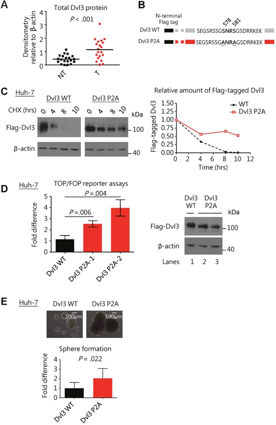
(A) Scatter plot showing a summary of Dvl3 protein expression in 20 paired HCC (T) and corresponding non-tumorous tissues (NT) as analyzed by Western blot densitometry. (B) Schematic diagram showing mutation of serine residues 578 and 581 on N-terminal Flag-tagged Dvl3. (C) The NP-Dvl3 mutant showed sustained protein stability at different time points as compared to the WT upon treatment with cycloheximide (CHX) at 10 ug/ml in Huh-7. (D) The NP-Dvl3 mutant was more active than WT Dvl3 in Huh-7. To ensure normalization of the amount of Dvl3 protein (right) to allow comparison in TOP/FOP reporter assays, DNA constructs were transfected at the following amounts: 2.5 μg of Dvl3 WT, 0.875 μg and 1.0 μg of Dvl3 P2A for lanes 1, 2, and 3, respectively. (E) The NP-Dvl3 mutant promoted greater sphere forming ability than WT Dvl3 in Huh-7. All in vitro experiments were carried out in at least 3 independent trials and the values are represented as mean ± SD.