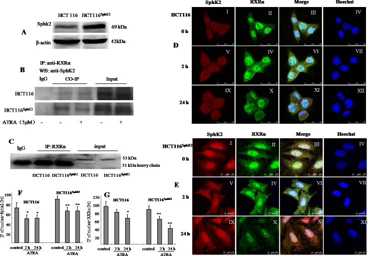
Figure 1. Sphk2 and RXRα are expressed in overlapping subcellular compartments in HCT-116 and HCT-116Sphk2 cells.
(A) Western blot of SphK2 in HCT-116 and HCT-116Sphk2 cells. (B) Western blot of co-immunoprecipitated SphK2 and RXRα in HCT-116 or HCT-116Sphk2 treated with or without ATRA for 6 h. HCT-116Sphk2 cells showed a strong immunoprecipitate band expressing RXRα contains SphK2. ATRA induced the degradation of RXRα in HCT-116Sphk2 cells, showing weaker immunoprecipitate. (C) A study to confirm the efficacy of Co-IP in which a primary antibody was assessed for its ability to pull out itself vs. normal IgG of the same species which served as the corresponding control. (D) and (E) Immunofluorescent microsgraphs of the subcellular localization of Sphk2 and RXRα in HCT-116 (D) and HCT-116Sphk2 (E) cells exposed to vehicle (first row), ATRA for 2 h (second row) and ATRA for 24 h (third row). SphK2 staining is shown in the first column (red, I, V, and IX). RXRα staining is shown in the second column (green, II, IV and X). Nuclei are stained with Hoechst (fourth column, IV, VII and XII). A merge of the three channels is shown in the third column. (D) Endogenous SphK2 staining is found in the nucleus and cytoplasm in HCT-116 whereas RXRα staining is mainly in nucleus with some residual staining in the cytoplasm. (E) Transfected SphK2 mainly resides in the nucleus in HCT-116Sphk2 and colocalizes with SphK2 in the absence of ATRA (I). Following ATRA treatment RXRα staining increases in the cytoplasm (2 h, IV) and then disappears in the cytoplasm (24 h, X). (F) and (G) Immunofluorescence (IF) of nuclear Sphk2 (%) and RXRα (%) were quantitative performed using ImageJ (NIH, http://rsb.info.nih.gov/ij/) and Open View software. Threshold subtraction and measurements of nuclear IF were done in a mask created according to Hoechst staining using NIH Image J. Bars represent means ± S.D. of 3 times. *, p < 0.05 vs. vehicle control.

