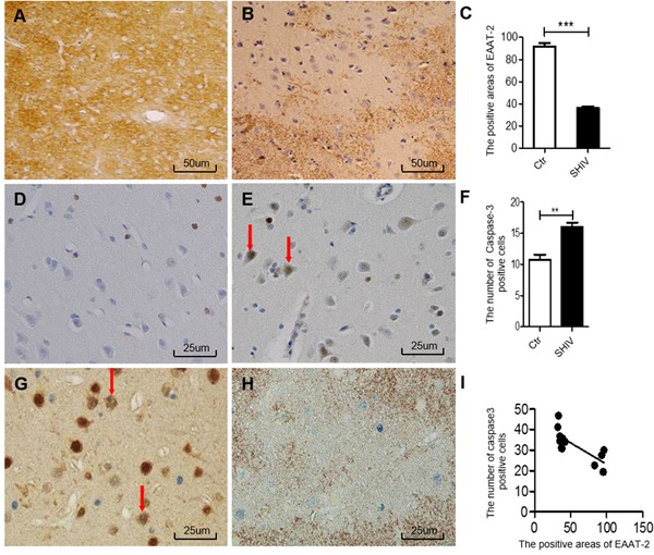
Figure 1. Association between decreased EAAT-2 and neuronal apoptosis in the cerebral cortex of SHIV-infected macaques.

A.-B. Decreased EAAT-2 expression in the cerebral cortex of SHIV-infected macaques (right) and controls (left). C. Statistical analysis of A and B (***P < 0.001). D.-E. The number of cleaved-caspase-3-positive cells in the cerebral cortex of SHIV-infected macaques (right) is increased compared with controls (left). F. Statistical analysis of D and E (**P < 0.01). G. Neuronal apoptosis in the cerebral cortex of SHIV-infected macaques as shown by NeuN (brown) and cleaved-caspase-3 (blue) double immunohistochemical staining. H. Neuronal apoptosis in areas with decreased EAAT-2 expression according to cleaved-caspase-3 (blue) and EAAT-2 (brown) double immunohistochemical staining. I. Statistical analysis of H demonstrates that areas with EAAT-2 expression correlate negatively with the number of cleaved-caspase 3-positive cells (*P < 0.05 and R2 = 0.5861). Ctr: uninfected group; SHIV: SHIV-infected group.
