Figure 5.
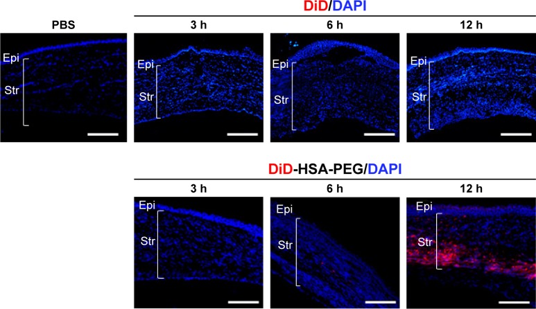
Intracorneal distribution of DiD-HSA-PEG nanoparticles and DiD after subconjunctival injection into alkali burn-injured rats.
Notes: Representative fluorescence microscopic images show central regions of corneas harvested 3, 6, or 24 h after subconjunctival injection of DiD-HSA-PEG nanoparticles, DiD, or PBS into alkali burn-injured rats. Fluorescent DiD (red) was used a model hydrophobic drug similar to apatinib. Cell nuclei were stained with DAPI (blue). Scale bars =100 µm.
Abbreviations: DiD, 1,1′-dioctadecyl-3,3,3′,3′-tetramethylindodicarbocyanine perchlorate; DiD-HSA-PEG, DiD-loaded human serum albumin-conjugated polyethylene glycol; DAPI, 4′,6-diamidino-2-phenylindole; PBS, phosphate-buffered saline; Epi, corneal epithelium; Str, stroma.

