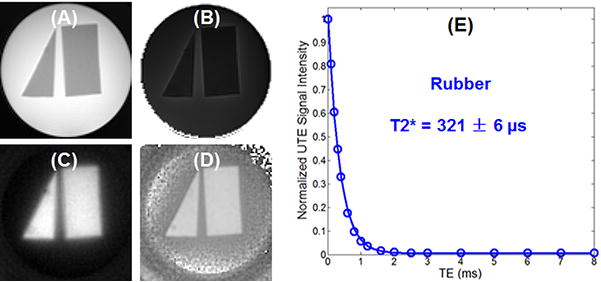
Figure 5.

shows both UTE and IR-UTE images of the rubber phantom. Excellent suppression of the agarose gel and selective magnitude and phase imaging of the rubber were achieved simultaneously using the IR-UTE technique. The rubber signal showed a single-component decay behavior with a short T2* of 321 ± 6 μs, which is slightly longer than T2* of protons in myelin lipid and myelin basic protein.
