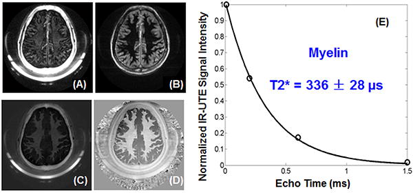
Figure 6.

IR-UTE imaging of the brain of a 38-year-old male volunteer with a TE of 8 μs (A) and 4.4 ms (B). Subtraction of the second echo from the 1st echo provides high contrast for myelin (C). Adaptive phase reconstruction of the phased array images provides high phase contrast (D). The weak signal from the white matter shows a fast decay with a short T2* of 336 ± 28 μs (E), suggesting WML being suppressed and myelin being detected.
