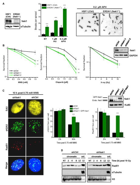
Figure 1. Nek1 Functions during the DNA Damage Response and Serves to Maintain Genomic Stability.
(A) Chromosome spreads of human fibroblasts. Chromatid breaks were analyzed in Nek1-deficient (ERDA1) and control (HSF1) cells both spontaneously (NT, not treated) and after a 20-hr exposure to HU or APH. Mean ± SEM (n = 3).
(B) Clonogenic survival of Nek1-deficient cells. Two independent shNek1 HeLa cell clones were generated by genomic shRNA insertion. Non-silencing shRNA was used as a control. DNA damage was induced by MMS (for 1 hr), olaparib (permanent), or X-rays. Mean ± SEM (n = 3).
(C) γH2AX and Rad51 foci in Nek1-depleted and GFP-Nek1-complemented cells. Asynchronous cells were co-treated with MMS and EdU for 1 hr. γH2AX and Rad51 foci were enumerated in EdU-positive cells (Figure S1B). Mean ± SEM (n = 3); spontaneous foci were subtracted.
(D) Chromatin fraction of Rad51 in Nek1-depleted cells. Synchronized cells were X-irradiated in G2 (Figure S1C) and chromatin fractions were analyzed for Rad51 by immunoblotting. H3 and αTubulin signals demonstrate the efficiency of chromatin fractionation (sol., soluble fraction).

