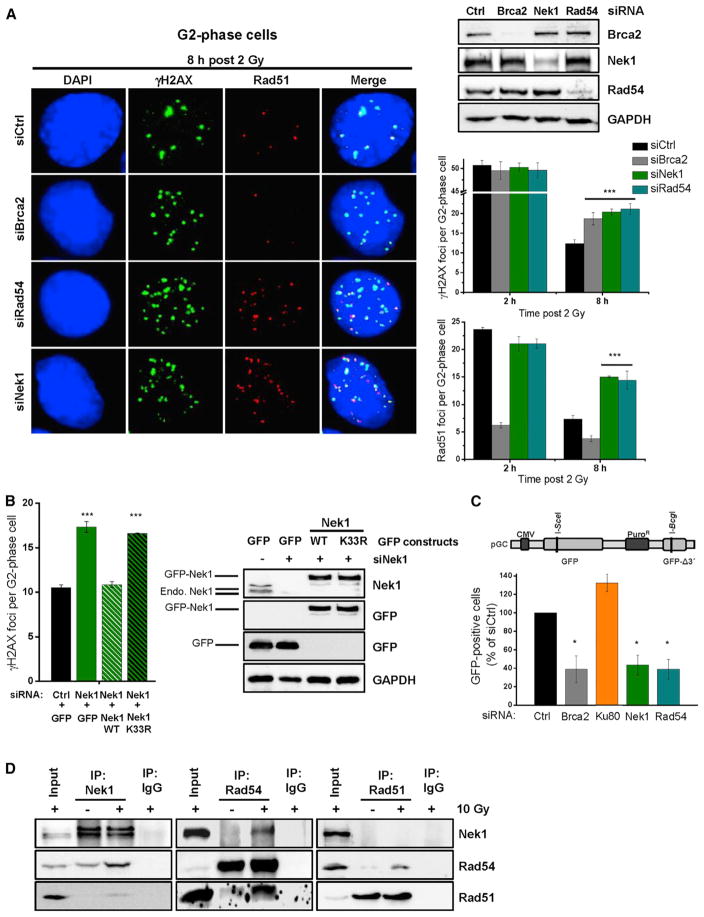
Figure 2. Nek1 Functions during DSB Repair by HR and Interacts with Rad54.
(A) γH2AX and Rad51 foci in Nek1-, Rad54-, and Brca2-depleted cells. HeLa cells were treated with siRNAs, EdU labeled, and X-irradiated. γH2AX and Rad51 foci were analyzed in EdU-negative G2-phase cells (Figure S2A). Mean ± SEM (n = 3); spontaneous foci were subtracted.
(B) γH2AX foci in catalytically deficient Nek1 cells. HeLa cells were treated with siNek1, transfected with siRNA resistant plasmids, X-irradiated, and γH2AX foci were enumerated 8 hr post 2 Gy in G2-phase cells identified as in (A). Mean ± SEM (n = 3); spontaneous foci were subtracted.
(C) GFP-based HR reporter assay with Nek1-, Rad54-, Brca2-, and Ku80-depleted cells. HeLa pGC cells were treated with siRNAs and transfected with an I-SceI plasmid. The number of GFP-positive cells was analyzed by IF microscopy. Mean ± SEM (n = 4).
(D) Physical interaction between Nek1, Rad54, and Rad51 in HeLa cells. Proteins were immunoprecipitated from nuclear cell extracts at 5 hr post irradiation and interactions were tested by immunoblotting.

