
Figure 2.
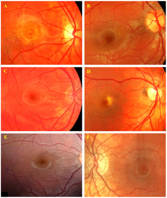
Fundus images of Benign Yellow Dot Maculopathy Subjects. A – Familial benign yellow dot maculopathy, Subject 11; B – Sporadic benign yellow dot maculopathy, fine, discrete, dots, Subject 24; C – Sporadic benign yellow dot maculopathy, concentrated in the nasal parafoveal region, Subject 26; D – Sporadic benign yellow dot maculopathy, confluent, Subject 28; E - Sporadic benign yellow dot maculopathy, Subject 34; F – Familial benign yellow dot maculopathy, Subject 19