Figure 3.

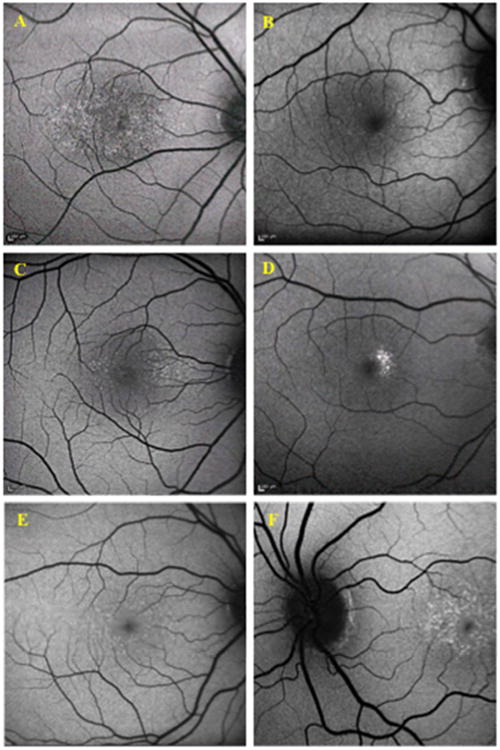
Fundus autofluorescence of Benign Yellow Dot Maculopathy Subjects. The yellow dots show hyperautofluorescence on fundus autofluorescence imaging. A –Subject 11; B –Subject 24; C –Subject 26; D – Subject 28; E – Subject 34; F – Subject 19

Fundus autofluorescence of Benign Yellow Dot Maculopathy Subjects. The yellow dots show hyperautofluorescence on fundus autofluorescence imaging. A –Subject 11; B –Subject 24; C –Subject 26; D – Subject 28; E – Subject 34; F – Subject 19