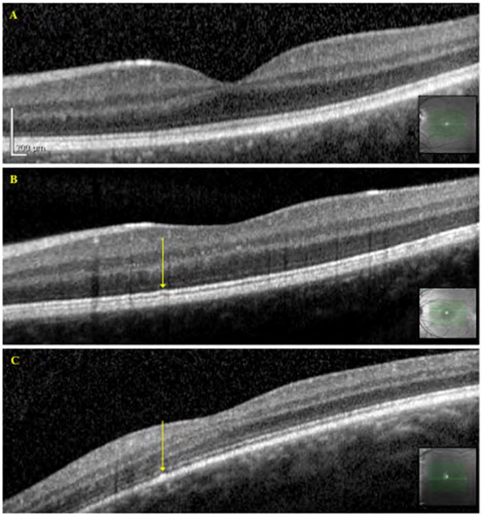
Figure 4.

OCT images of Benign Yellow Dot Maculopathy Subjects. A – Normal OCT, Subject 24; B – Slight irregularity of the inner segment ellipsoid band, indicated by the arrow, Subject 26; C – Slight irregularity of the RPE layer, indicated by the arrow, Subject 11. Arrows correspond to the location of the dots as identified from the infrared reflectance images obtained during fundus autofluorescence image acquisition
