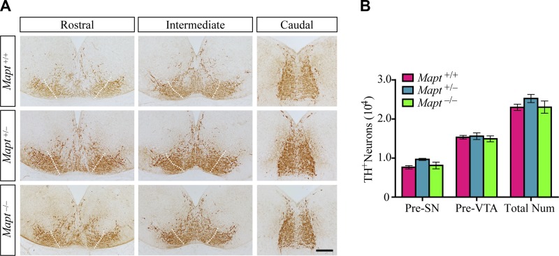
Figure 1.
Normal mDAN morphology and number in tau-deficient mouse embryos at E14.5. A) TH immunohistochemistry of midbrains of E14.5 Mapt+/+, Mapt+/−, and Mapt−/− mice. Representative coronal sections of midbrain, rostral to caudal, are shown, with white dotted lines demarcating the presumptive (Pre) boundary between the SN and VTA. The lateral TH+ area is considered to be Pre-SN, whereas the intermediate is Pre-VTA. Scale bar, 100 μm. B) The number of TH+ neurons in the Pre-SN, Pre-VTA, and total midbrain of E14.5 mice (n = 5 for each genotype vs. control littermates). Data are means ± sem.

